Stochastic electrotransport selectively enhances the transport of highly electromobile molecules [full article]

Sung-Yon Kim, Jae Hun Cho, Evan Murray, Naveed Bakh, Heejin Choi, Kimberly Ohn, Sara Vassallo, Luzdary Ruelas, Austin Hubbert, Meg McCue, Philipp Keller and Kwanghun Chung. Stochastic electrotransport selectively enhances the transport of highly electromobile molecules, PNAS, 2015 Nov 17: 112(46): E6274-83. doi: 10.1073/pnas.1510133112. Epub 2015 Nov 2. PubMed PMID: 26578787; PubMed Central PMCID: PMC4655572.


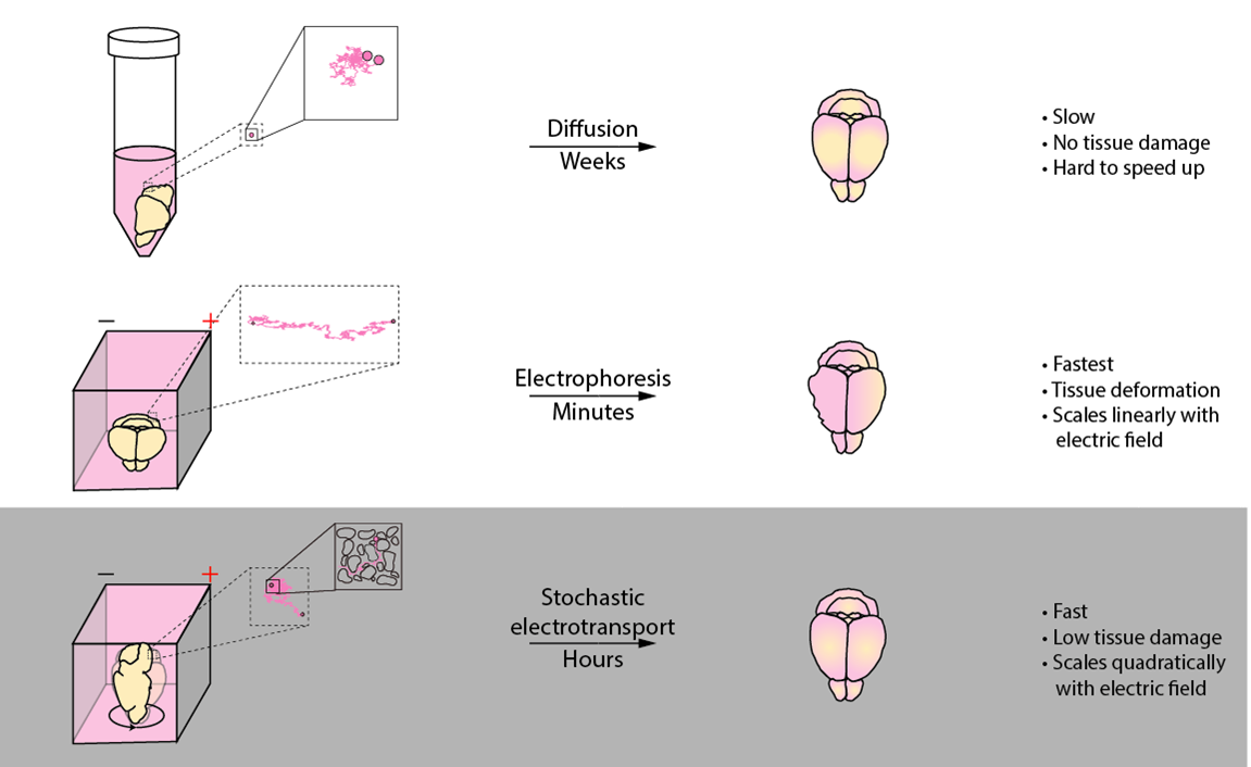
Stochastic electrotransport is a transport enhancement method designed specifically for biological tissues. Like electrophoresis, it uses electric fields to drive electromobile molecules into the tissue. But unlike electrophoresis, it does not cause damage to the tissue. This allows you to quickly process tissues without destroying them.


How does it work? Stochastic electrotransport uses a rotational electric field. This rotational electric field creates dispersion - a diffusion-like transport.

The illustration above compares stochastic electrotransport to electrophoresis (static electric field) and diffusion (no electric field).

Imagine you're a particle inside of a tissue. You are the magenta dot in the middle of the top left panel. To you, the microscopic structure in the tissue looks like a maze - represented by random black spots that you can't move through. Naturally, you want to move around and explore - this is called diffusion. But diffusion is a slow process and you don't get that far. An electric field could help you, but only in a particular direction - this causes stress to both you and the structure around you, which also feel the electric field. A rotational electric field, on the other hand, pushes you in all direction. This helps you spread out.

In the article, we show, using experiments and simulations, that this enhancement is quadratic with respect to the electromobility and the electric field and linear with respect to the rotation period, if the electric field is large enough. This means that 10X electric field (loosely equivalent to the voltage) would result in 100X transport. Likewise, 10X increase in electromobility (for a protein, that means higher deviation from neutral pH) would result in 100X transport. The dependence on rotation period is linear, so a 10X increase in rotation period (say, 1 minute to 10 minutes per rotation) would result in 10X transport. The moral of the story is, we want to apply the highest voltage possible and the lowest rotation rate possible that does not damage the tissue.

Application: Clearing

One application for stochastic electrotransport is tissue clearing. We use the detergent SDS to remove light-scattering lipids from the tissue. This process can be sped up by speeding up the transport of SDS into and lipids out of the tissue. In a higher level of abstraction, it is simply a process where electromobile particles are transported into and out of tissues, and we can do this quickly and nondestructively using stochastic electrotransport.

We developed a specialized device to carry out this task:

The device is simpler than it seems. The key is that the tissue is placed between the electrodes. This tissue is then rotated to create a rotational electric field. With the right conditions, you can clear tissues quite rapidly, without harming the tissue at all.

Picture6.png


Clearing Protocol

These are the materials that you will need:

Name Vendor Catalogue Number
Boric acid Sigma B7901
Lithium Hydroxide Sigma 254274
Sodium Dodecyl Sulfate Sigma L3771
Spectra/Por 1 Spectrum Labs 132655
Custom constructed device N/A N/A

Granted, the hardest part will be constructing your device. We've included a blueprint for the device in the supplementary materials of the paper. We're also working with a manufacturer to produce these devices, so stay tuned.

You'd need to make the following solutions:

  • Inner: 200 mM SDS, 10 mM Lithium Hydroxide, 20 mM Boric Acid
  • Outer: 10 mM SDS, 10 mM Lithium Hydroxide, 20 mM Boric Acid

There are two different solutions: Inner and Outer. The Inner solution has the optimum concentration of SDS necessary for rapid tissue clearing. The Outer solution has just enough SDS to maintain micellar equilibrium with the Inner solution SDS. Having these two solutions significantly decreases SDS degradation - meaning less maintenance and lower costs.

Once everything is ready to go, pour the Inner solution into the Inner reservoir and the Outer solution into the Outer reservoir. Put the brain inside of the rotating "sample chamber" (which is just a cylindrical nylon mesh to make sure the tissue doesn't float away). Then, apply the maximum voltage you can across the electrodes that does not cause (1) bubble formation in the tissue, (2) runaway Joule heating (i.e. temperate keeps rising and rising), or (3) damage to the device (the membranes, plastic parts, or the electrodes). There isn't a magical voltage for this - it's all trial and error. It depends on your device geometry (namely, distance between the electrodes) and your tissue. This is one of the reasons why we're trying to produce standardized devices with well-characterized properties and easy-to-follow protocols.

Application: Labeling

Another application for stochastic electrotransport is labeling (aka staining). Labeling is much more complex than clearing and can depend on many factors. It's also much harder to figure out the right conditions for labeling. But again, labeling, in abstract terms, is simply transporting electromobile particles into the tissue, where they will bind to their targets. And stochastic electrotransport can speed this process up.

There is another specialized device for this:

The key concept - rotating the tissue with respect to parallel electrodes - is the same. But this also has a nanoporous membrane to confine the probes (antibodies, for example) within a small volume next to the tissue.

With the right conditions, you can get labeling like this:

And like these:

Labeling Protocol

These are the materials that you will need:

Name Vendor Catalogue Number
Boric acid Sigma B7901
Lithium Hydroxide Sigma 254274
Molecular Probes Any Any
Spectra/Por 1 Spectrum Labs 132655
Custom constructed device N/A N/A

Again, the hardest part will be constructing your device. We've included a blueprint for the device in the supplementary materials of the paper. We're also working with a manufacturer to produce these devices, so stay tuned.

You'd need to make the following solutions:

  • Electrophoresis: 50 mM Lithium Hydroxide, 25 mM Boric Acid
  • Staining: 50 mM Lithium Hydroxide, 25 mM Boric Acid, 1% Triton-X 100, 0.02% Sodium Azide

Staining solution is the same as Electrophoresis solution, except that it contains 1% Triton-X 100 to minimize the adsorption of molecular probes onto the membranes and to permeabilize the tissue as well as sodium azide to prevent bacterial growth.

Before you begin labeling, you need to make sure the tissue contains no SDS, which interferes with labeling. To do that, we recommend you wash your tissue three times over three days in the Staining solution.

Once you have everything, pour the Electrophoresis solution into the Electrophoresis reservoir. Then, add enough Staining solution into the sample chamber to completely immerse the tissue. Pipet mix molecular probes into the Staining solution. Then, apply the maximum voltage you can across the electrodes that does not cause (1) bubble formation in the tissue, (2) runaway Joule heating (i.e. temperate keeps rising and rising), or (3) damage to the device (the membranes, plastic parts, or the electrodes). This is similar to clearing above. After about 24 hours (to make sure the labeling is done), take out the tissue and incubate it in your desired optical clearing solution. We use what we call PROTOS. You can find details about PROTOS in an upcoming post.


How to orient samples for optimal Stochastic Electrotransport clearing

Stochastic electrotransport (SE) is a novel method for enhancing the transport of electromobile molecules.  SE clearing utilizes rotational electrical field to achieve high speed clearing of biological tissues without tissue damage. Clearing or clarification refers to the removal of lipids from tissues samples in order to improve its optical transparency for imaging and chemical permeability for labeling.

Lipid removal is achieved by the use of surfactant micelles, and the clearing speed of a biological tissue is dependent on how fast the surfactant micelles can travel through the sample. These surfactant molecules are electrically charged and applying electric field accelerates the transport of the surfactant-lipid micelles complexes, enabling much faster clearing.

The SE clearing adjusts parameters such as the strength of the electric field to improve the effective diffusivity of detergent micelles and thereby significantly reduce the total diffusion time compared to that of a simple passive clearing scheme.

Total diffusion time scale (T) is dependent on the diffusion length (l) and the diffusivity of the micelles (D<sup>eff</sup>)

However, even with improved diffusivity, the clearing time’s quadratic dependence on the diffusion length remains. Thicker tissues will always take much longer to clear with the diffusion-based methodology. For SE clearing, electric field mediated diffusion occurs mostly along the direction of the electric field. This means that when clearing non-spherical samples, the orientation of the tissue samples undergoing SE clearing can have a significant impact on the total clearing time. As such, utmost consideration should be given to minimize the diffusion length that surfactant micelles will travel when loading the samples into the machine. Consider the following diagram:

diagram 1.PNG

The diagram shows two different orientations of a typical mouse hemisphere sample in a SE clearing chamber. The maximum diffusion length along the direction of electrical field for the left sample is almost 3 times longer than that of the right sample. Even with a same sample, the maximum diffusion length is 3 fold different. Because of the aforementioned quadratic dependence on diffusion length, the right sample can take 9 times or even longer to fully clear. Given that the time it takes to fully clear a correctly positioned mouse hemisphere can be up to 3 days, improperly positioned sample can remain uncleared for weeks.  

Optimal sample orientation will allow the tissues to be cleared quickly and without damage. Such orientations can be achieved by using nylon meshes that can help hold the samples in place.


Discussion

We want this to be a two-way discussion. Please post any questions or comments below.