Sign In
My Account
SHIELD
Introduction
Protocol
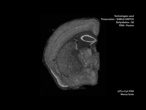
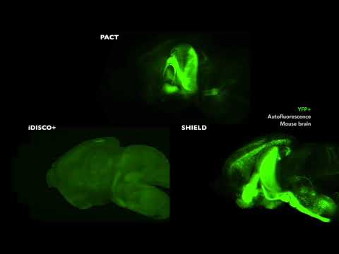
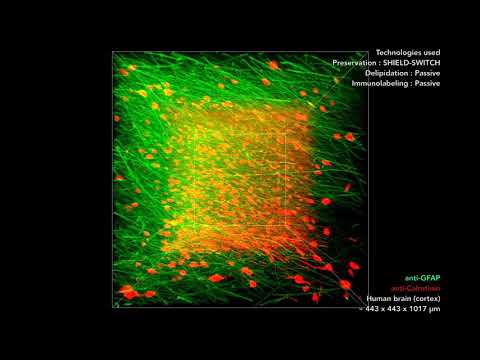
Video Gallery
Discussion
CLARITY
Introduction
Protocol
Stochastic Electrotransport
Introduction
How it works
Clearing
Labeling
Sample Orientation
Discussion
SWITCH
Introduction
How it works
Fixation
Inactivation and Clearing
Labeling
Video Gallery
Discussion
MAP
Introduction
Video Gallery
Probes
Chung Lab
Chung Lab Resources
Sign In
My Account
SHIELD
Introduction
Protocol
Video Gallery
Discussion
CLARITY
Introduction
Protocol
Stochastic Electrotransport
Introduction
How it works
Clearing
Labeling
Sample Orientation
Discussion
SWITCH
Introduction
How it works
Fixation
Inactivation and Clearing
Labeling
Video Gallery
Discussion
MAP
Introduction
Video Gallery
Probes
Chung Lab
1
2
3
4
5
6
7
8
9
10
11
Previous
Next